Dynamic needle tip positioning
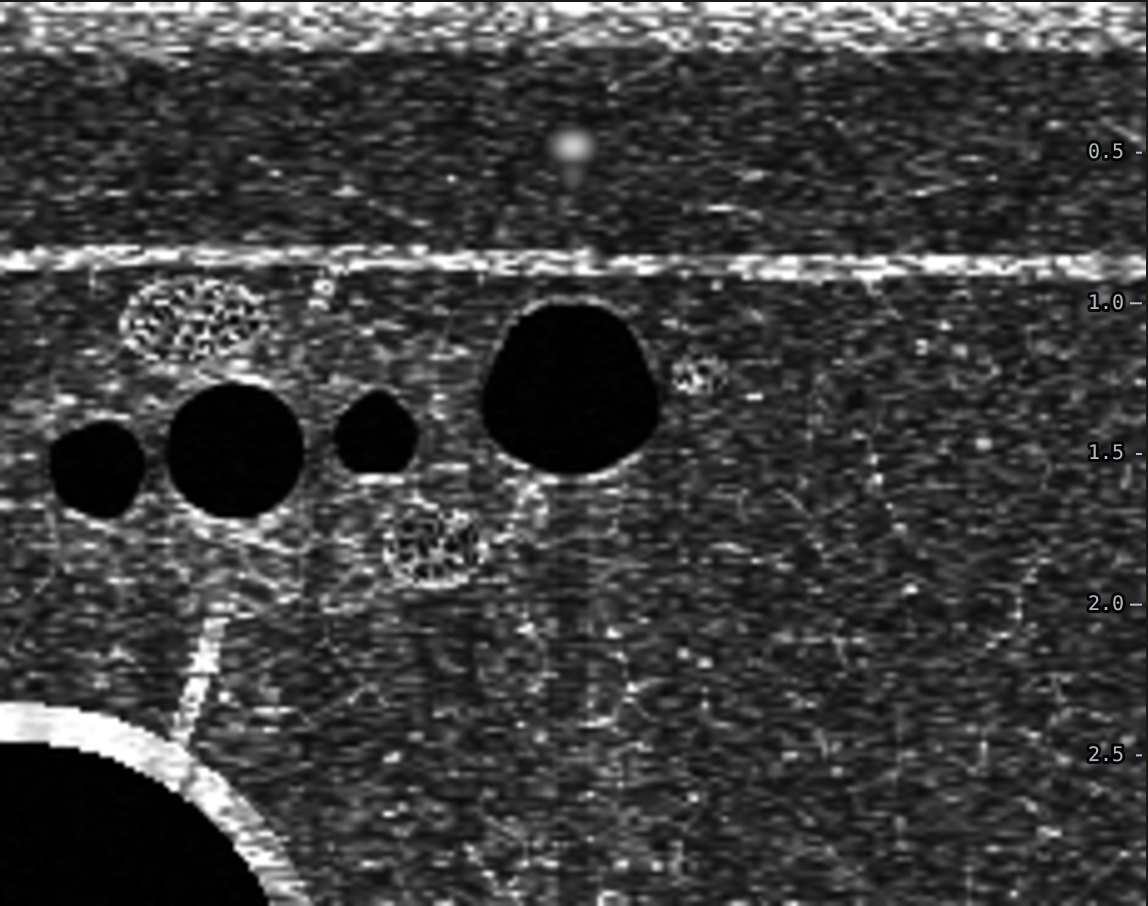
In a short-axis ultrasound view the needle shows as a single bright dot. The problem is that the shaft and the tip look the same. Dynamic needle tip positioning is how you make sure the dot you are watching is the tip.
Why the dot can lie
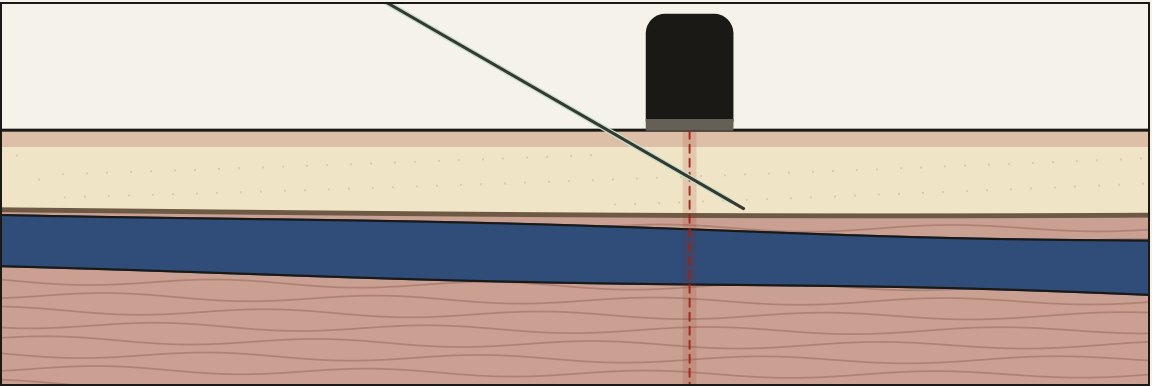
The ultrasound beam is a thin slice, about a millimetre thick. Wherever the needle crosses that slice, you see a dot. If the tip has already passed through the slice, the dot is part of the shaft and the tip is somewhere deeper, out of view. It can be through the back wall or in the artery while the screen still shows a dot sitting nicely above the vein.
The technique
- Puncture close to the probe so the tip shows up shallow.
- When you see the dot, slide the probe 1 mm away from the needle, toward the shoulder, until the dot just disappears. The beam is now just ahead of the tip.
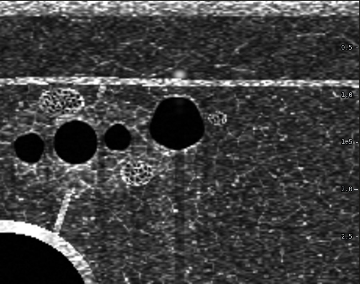
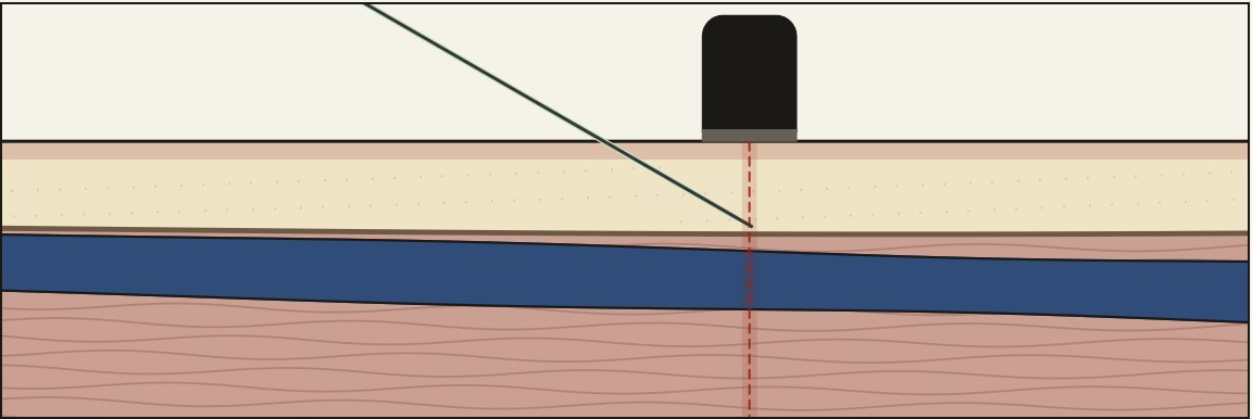
- Advance the needle until the dot reappears. Now the dot is the tip.
- Repeat in small steps until the tip is in the middle of the vein. A dot centered in the lumen is called the target sign.
- Flatten the needle and do two or three more cycles inside the vein so the catheter follows the tip in.
What the evidence shows
The technique was described by Clemmesen and colleagues in a blinded phantom trial with ultrasound novices (Clemmesen 2012). A 2023 network meta-analysis of pediatric randomized trials found higher first-attempt success with it than with palpation (RR 1.67) or with short-axis ultrasound without it (RR 1.43) (Takeshita 2023). The adult evidence is thinner, but the geometry is the same.
Tilting instead of sliding
Some people fan the probe toward the shoulder instead of sliding it. That moves the deep part of the beam ahead of the tip the same way. Sliding keeps the beam straight up and down, which is easier to judge.
Practice it on a simulated arm. The Ultrasound IV Trainer lets you scan, compress, pick a catheter and walk the needle tip into the vein in short or long axis, then shows a side view of where the tip and catheter really ended up.
Related guides
References
- Clemmesen L, Knudsen L, Sloth E, Bendtsen T. Dynamic needle tip positioning: ultrasound guidance for peripheral vascular access. A randomized, controlled and blinded study in phantoms performed by ultrasound novices. Ultraschall Med. 2012;33(7):E321-E325.
- Takeshita J, Nakayama Y, Tachibana K, Nakajima Y, Shime N. Ultrasound-guided short-axis out-of-plane approach with or without dynamic needle-tip positioning for peripheral venous catheterization in pediatric patients: a systematic review with network meta-analysis. J Cardiothorac Vasc Anesth. 2023.
- Blanco P. Ultrasound-guided peripheral venous cannulation in critically ill patients: a practical guideline. Ultrasound J. 2019;11:27.