Short axis vs long axis for ultrasound-guided IVs
There are two ways to line up the probe for an ultrasound-guided IV. Across the vein, it is short axis and the needle comes in out of plane. Along the vein, it is long axis and the needle is in plane. Each has a different problem to solve.
Short axis (out of plane)
The vein is a black circle and the needle is a dot. You see the vein's neighbours, the artery and nerve, at the same time, and it is easy to center on the vein. The catch is that the dot can be the shaft instead of the tip, so you have to follow the tip with dynamic needle tip positioning.
Long axis (in plane)
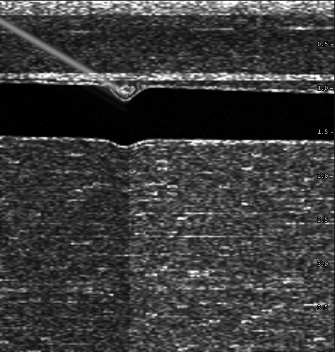
The vein is a horizontal tube and the whole needle is a bright line with the tip at its end. You can see exactly how deep the tip is. The catch is that the beam is only about a millimetre thick. A small sideways drift of the needle or the probe loses part or all of the line, and you cannot see the artery or nerve beside the vein.
The needle also looks brighter when it is flatter. A steep needle reflects its echo away from the probe, so in long axis a faint line often just means the needle is steep.
What the trials found
For peripheral IVs, a meta-analysis of three trials found higher success with short axis (97.7% vs 89.1%, OR 5.35), no clear difference in needle passes, and mixed results for time (Gottlieb 2018). That is a small evidence base. Long axis is still useful for straight, larger veins and as a check on tip depth.
Practice it on a simulated arm. The Ultrasound IV Trainer lets you scan, compress, pick a catheter and walk the needle tip into the vein in short or long axis, then shows a side view of where the tip and catheter really ended up.
Related guides
References
- Gottlieb M, Holladay D, Peksa GD. Comparison of short- vs long-axis technique for ultrasound-guided peripheral line placement: a systematic review and meta-analysis. Cureus. 2018;10(5):e2718.
- Clemmesen L, Knudsen L, Sloth E, Bendtsen T. Dynamic needle tip positioning: ultrasound guidance for peripheral vascular access. A randomized, controlled and blinded study in phantoms performed by ultrasound novices. Ultraschall Med. 2012;33(7):E321-E325.
- Blanco P. Ultrasound-guided peripheral venous cannulation in critically ill patients: a practical guideline. Ultrasound J. 2019;11:27.