Finding the vein on ultrasound
Most ultrasound-guided IV failures start with the vein choice. Here is how to find the vein, prove it is a vein, and decide whether it is worth the attempt.
Where to look
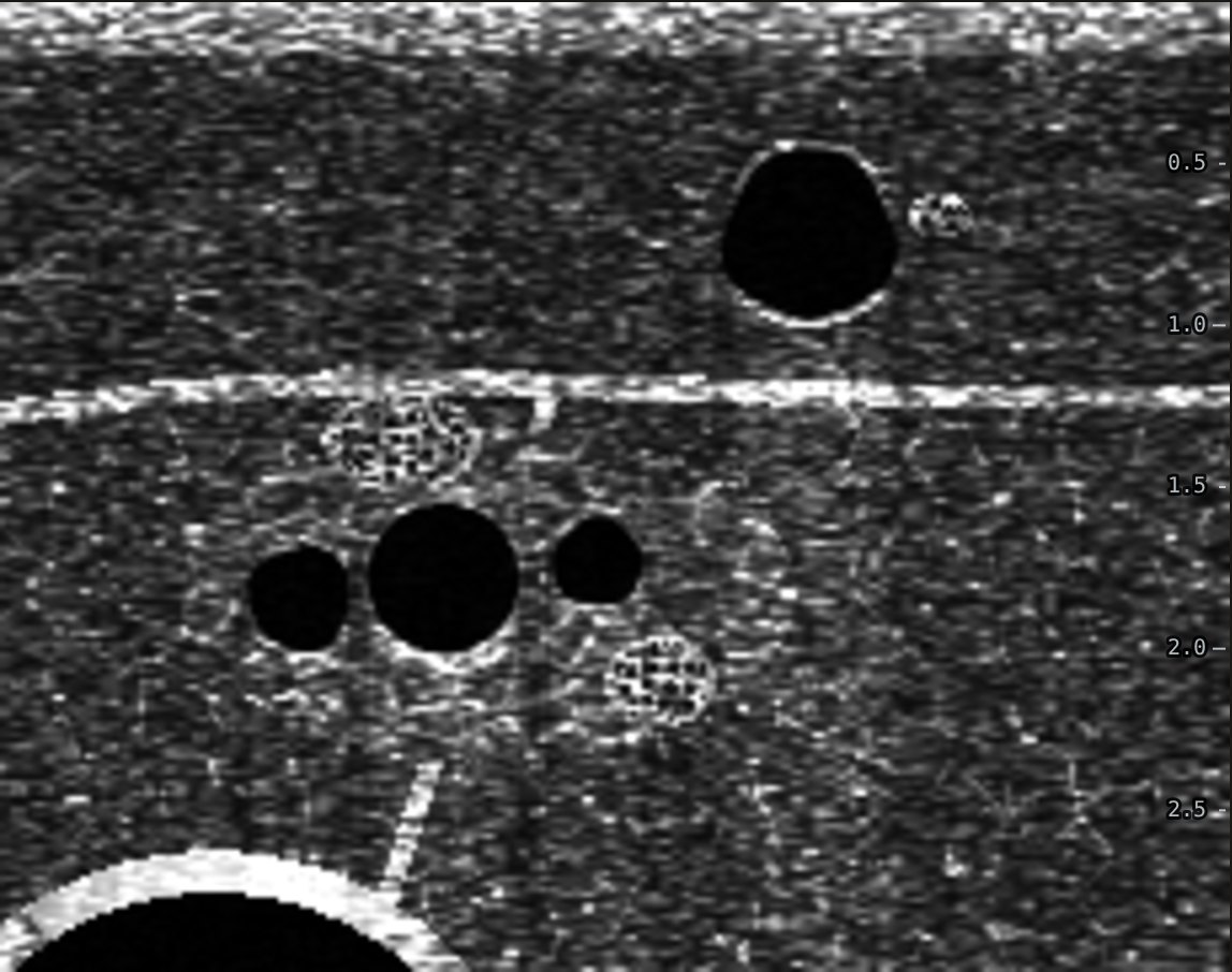
Start on the medial upper arm, just above the elbow crease, probe across the arm. The basilic vein lies in the subcutaneous fat. Follow it up the arm: it gets deeper and eventually dives under the fascia. Deeper still, the brachial artery runs with two paired brachial veins and the median nerve, which looks like a small honeycomb.
Vein or artery
- Compression. Gentle pressure flattens a vein completely. An artery stays round.
- Pulsation. The artery pulses and has a thicker, brighter wall.
- Color Doppler. In short axis, tilt the probe a little to get color. Arterial flow pulses and often aliases; venous flow is slow and steady.
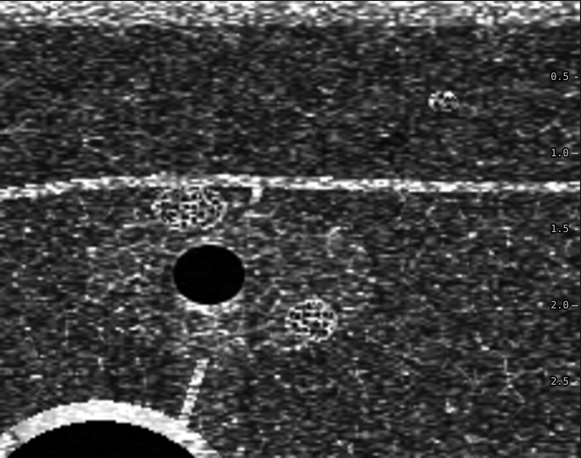
A vein that will not flatten, or has gray material inside it, may be clotted. Choose another segment or another vein.
Size and depth
Bigger is better: each extra 1 mm of diameter raised the odds of success, and depth did not matter until 1.6 cm, beyond which no vein was cannulated (Panebianco 2009). Veins at least 4 mm wide and 0.3 to 1.5 cm deep had the highest success (Witting 2010). If two veins are both under 1.6 cm, take the wider one even if it is deeper, and pick a catheter long enough for it.
What to avoid
- Brachial veins hugging the artery and nerve, when the basilic is usable.
- Veins that bend sharply or have a valve right where you plan to thread.
- Small superficial veins that look easy but are under 3 mm wide.
Practice it on a simulated arm. The Ultrasound IV Trainer lets you scan, compress, pick a catheter and walk the needle tip into the vein in short or long axis, then shows a side view of where the tip and catheter really ended up.
Related guides
References
- Panebianco NL, Fredette JM, Szyld D, et al. What you see (sonographically) is what you get: vein and patient characteristics associated with successful ultrasound-guided peripheral intravenous placement in patients with difficult access. Acad Emerg Med. 2009;16(12):1298-1303.
- Witting MD, Schenkel SM, Lawner BJ, Euerle BD. Effects of vein width and depth on ultrasound-guided peripheral intravenous success rates. J Emerg Med. 2010;39(1):70-75.
- Blanco P. Ultrasound-guided peripheral venous cannulation in critically ill patients: a practical guideline. Ultrasound J. 2019;11:27.