IV catheter length for ultrasound-guided IVs
Ultrasound lets you reach deeper veins, but every millimetre between the skin and the vein wall is catheter that is not in the vein. Pick the length before you puncture.
The 65% rule
In ultrasound-guided IVs, every catheter with less than 30% of its length inside the vein failed, about a third failed with 30 to 64%, and none failed with 65% or more. Each extra 5% inside the vein cut the hazard of failure by 29% (Pandurangadu 2018).
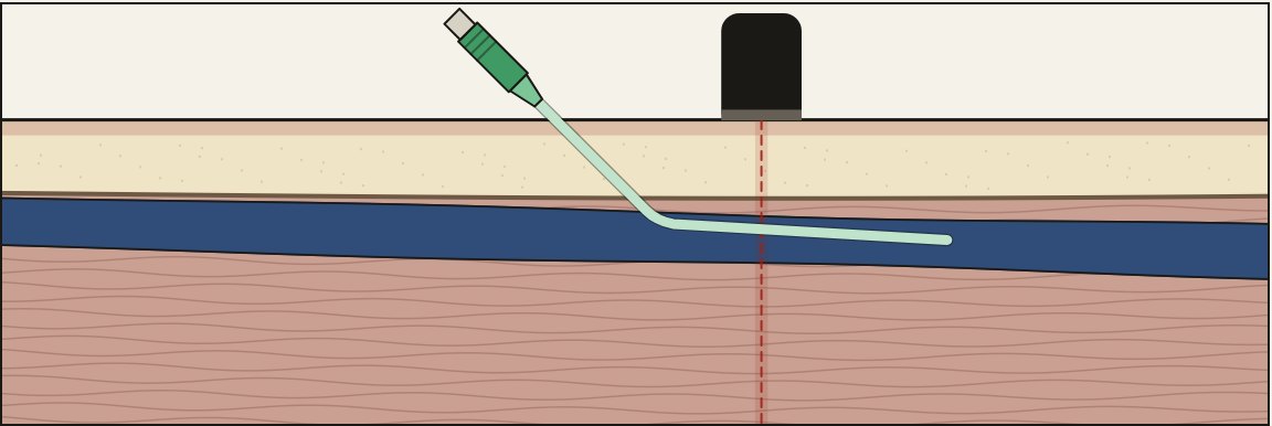
Estimate the track
Catheter needed ≈ track ÷ 0.35
At 45 degrees, sin is about 0.7, so the track is about 1.4 times the depth. A vein whose top wall is 1 cm deep has a track of about 1.4 cm, and keeping 65% inside needs a catheter of about 4 cm. A 1.25 inch (3.2 cm) catheter leaves only about 55% in the vein. A 1.88 inch (4.8 cm) catheter leaves about 70%.
| Top wall depth | Track at 45° | Shortest catheter for 65% |
|---|---|---|
| 0.5 cm | 0.7 cm | 2 cm (1.25 in works) |
| 1.0 cm | 1.4 cm | 4 cm (1.88 in) |
| 1.5 cm | 2.1 cm | 6 cm (2.5 in) |
Flattening the needle after you are in the vein does not shorten the track. The track is set where you go through the vein wall.
Long vs standard catheters
In a randomized trial of ultrasound-guided IVs, long catheters failed less often than standard ones (14% vs 45%), at the cost of a few extra minutes to place (Elia 2012).
Catheter width
A catheter that fills a large share of the vein raises the risk of clot and early failure. A common rule of thumb is to keep the catheter under about 45% of the vein's diameter, which mostly comes from PICC research. In a 3 mm vein, a 20G fits better than an 18G.
Practice it on a simulated arm. The Ultrasound IV Trainer lets you scan, compress, pick a catheter and walk the needle tip into the vein in short or long axis, then shows a side view of where the tip and catheter really ended up.
Related guides
References
- Pandurangadu AV, Tucker J, Brackney AR, Bahl A. Ultrasound-guided intravenous catheter survival impacted by amount of catheter residing in the vein. Emerg Med J. 2018;35(9):550-555.
- Elia F, Ferrari G, Molino P, et al. Standard-length catheters vs long catheters in ultrasound-guided peripheral vein cannulation. Am J Emerg Med. 2012;30(5):712-716.
- Blanco P. Ultrasound-guided peripheral venous cannulation in critically ill patients: a practical guideline. Ultrasound J. 2019;11:27.